How to Use Disposable Skin Stapler Safely in Veterinary Practice
-
March 16, 2026 11:15 AM PDT
Closing wounds quickly and safely is one of the most important responsibilities in veterinary medicine. Whether it is a routine general surgery, an urgent laceration repair, a complex orthopedic surgery, or a high-risk cesarean section, veterinarians rely on efficient tools to achieve precise and stable skin closure. Among these tools, the disposable skin stapler has become a preferred option in many clinics thanks to its speed, consistency and ease of use.
When applied correctly, skin staplers offer faster closure compared to traditional sutures, reduce anesthesia time, and minimize tissue handling. However, safe and effective disposable skin stapler use requires proper technique, understanding of staple mechanics and careful post-operative management. This detailed guide covers essential methods, best practices and clinical considerations for using skin staplers across various wound closure methods in animals.
Why Disposable Skin Staplers Are Used in Veterinary Procedures
Skin staplers are widely adopted in veterinary medicine because they help clinicians maintain wound precision while minimizing the time an animal spends under anesthesia. In trauma cases, rapid closure can be life-saving. In surgical settings, reduced manipulation of the skin lowers the risk of inflammation and speeds recovery.
Key benefits include:
-
Fast skin approximation, ideal for emergency and high-volume practices
-
Consistent staple spacing, ensuring reproducible results
-
Minimal tissue reaction, especially when compared with some suture materials
-
Reduced hand fatigue, which is valuable during long or repetitive procedures
1. Preparing the Wound: The Foundation of Safe Stapler Use
Before using a disposable skin stapler, wound preparation is the most important step. Even the highest-quality skin stapler cannot compensate for poor preparation.
Cleaning and Debridement
All debris, hair, necrotic tissue and contaminants must be removed. A clean wound bed reduces infection risk and ensures proper bonding between opposing skin edges.
Local Anesthesia
Even in cooperative animals, proper local anesthesia prevents discomfort and reduces movement during closure. Local blocks or regional anesthesia may be used depending on the wound location.
Achieving a Moisture-Controlled Field
The skin must be dry before stapling. Residual blood, antiseptics or fluid reduces staple grip and may lead to premature staple loosening.
Evaluating Tension and Edges
If skin edges cannot meet without tension, stapling is not recommended. High tension increases the risk of wound dehiscence. Alternative methods such as intradermal sutures or tension-relieving patterns may be needed before stapler use.
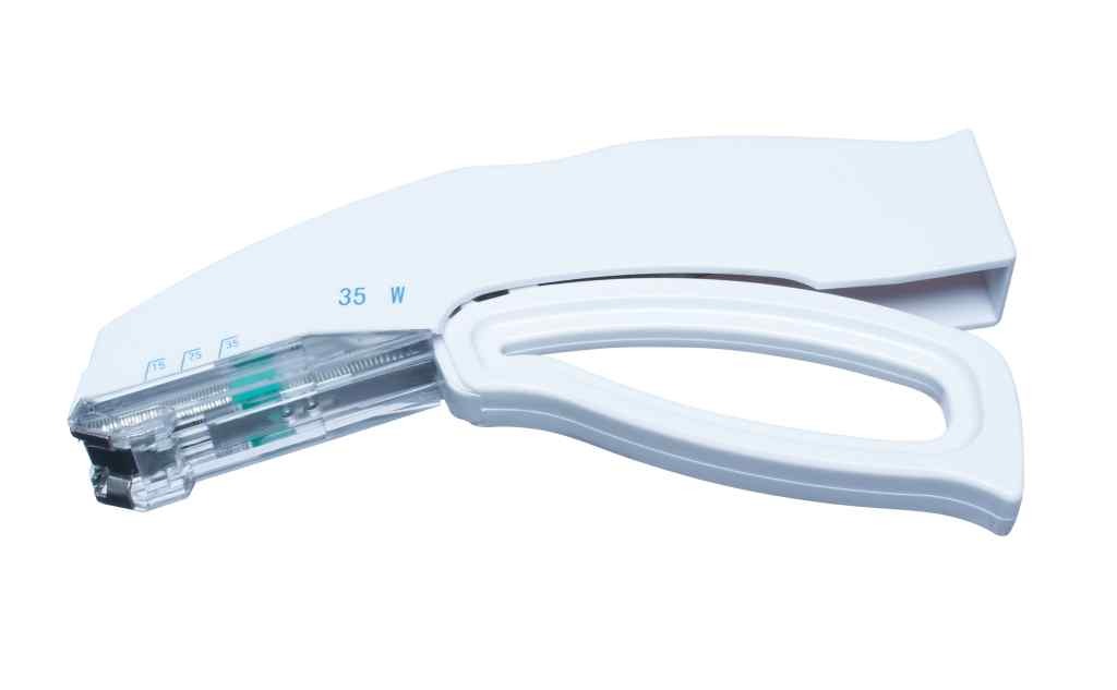
2. Understanding Stapler Loading and Positioning
Correct stapler loading and stapler positioning are essential for predictable and safe closure. While disposable staplers come preloaded, clinicians must still understand their internal mechanism.
How Disposable Skin Staplers Work
Most staplers deliver one staple per trigger squeeze and automatically advance the next one. The preloaded magazine improves efficiency and sterility, making them ideal for both routine and emergency use.
Stapler Positioning
Proper alignment ensures even and symmetrical closure:
-
The stapler should be held perpendicular to the skin.
-
The tips must rest lightly on the skin surface without compressing underlying tissue.
-
The skin edges should be perfectly everted before firing.
Eversion is critical because inverted or flat edges lead to poor cosmetic healing and increased scar formation.
Spacing Between Staples
Most disposable staplers place staples at optimal intervals automatically, but veterinarians must still monitor spacing and adjust based on:
-
Animal size
-
Skin thickness
-
Tension on the wound
Too-wide spacing reduces support while too-close spacing increases unnecessary trauma.
3. Technique for Safe and Effective Staple Application
A smooth technique produces consistent and stable closure across species.
Step-by-Step Application
-
Use forceps to carefully bring wound edges together, ensuring proper alignment and minimizing tension before applying each surgical skin staple.
-
Position the stapler jaws precisely along the wound line, maintaining even spacing to support accurate staple placement and consistent wound closure.
-
Ensure the skin edges are slightly everted before stapling, promoting better healing, reduced scarring, and stronger long-term wound edge stability.
-
Apply smooth, steady pressure to the stapler handle, creating a secure staple that holds tissues together without crushing surrounding skin.
-
After releasing the handle, inspect the staple to confirm correct formation, ensuring both legs penetrate evenly and secure the wound edges properly.
-
Advance along the wound, maintaining consistent spacing between staples, supporting uniform tension distribution and promoting predictable, high-quality wound healing outcomes.
The goal is a uniform row of staples that mirror each other and support the healing process.
Confirming Staple Shape
A properly placed staple resembles a rectangular "box" shape when viewed from above. If the legs are uneven or one leg fails to penetrate the skin, the staple must be removed and replaced immediately.
Staple Removal Indicator
Many staplers include a staple removal indicator on the packaging or instructions. This helps clinicians determine the optimal removal window based on animal species, anatomical location and degree of tension.
4. Using Skin Staplers in Specific Veterinary Procedures
Because animals vary widely in skin elasticity, tissue density and movement patterns, the correct stapler technique shifts slightly by procedure type.
Laceration Repair in Trauma Care
In emergency situations where speed matters, skin staplers allow rapid closure while minimizing contamination. However, if the wound is heavily contaminated or irregular, delayed closure or partial primary closure may be preferable.
Orthopedic Surgery
Animals receiving orthopedic repairs often have significant postoperative movement. Staples provide strong mechanical resistance along incision lines, reducing the risk of wound dehiscence.
Cesarean Section and Gynecological Surgeries
Staplers reduce closure time, lowering anesthesia risk for both the mother and offspring. They also reduce the manipulation of swollen or vascular abdominal skin.
Dermatological Procedures
After mass removal or biopsy, staplers create neat approximations and minimize tissue trauma. They are ideal for short procedures requiring clean, quick closure.
General Surgery
In routine procedures like abdominal exploratory surgeries, skin staplers are often chosen for their uniformity and speed. Internal layers are still closed with sutures, while the outer skin is stapled for efficiency.
5. Post-Stapling Care and Monitoring
Successful closure does not end at the moment of stapling. Post-stapling care ensures long-term healing and wound integrity.
Monitoring for Infection
Daily evaluation is essential, especially in active animals. Watch for:
-
Redness
-
Swelling
-
Discharge
-
Pain on palpation
Any signs of infection warrant re-evaluation of the wound and possibly early staple removal.
Wound Dressing
Light dressings may be applied in high-movement areas or for animals prone to licking. An Elizabethan collar is often recommended to prevent self-trauma.
Staple Removal
Stapler stitch removal is typically performed using a dedicated skin stapler remover, which gently bends the staple to disengage it. Removal times vary for each case:
-
10–14 days for most dogs and cats
-
Up to 21 days for high-tension areas or large breeds
The tissue should appear epithelialized and stable before removing staples. Veterinarians should be consulted for this.
6. Comparing Skin Staplers With Other Wound Closure Methods
Although this guide focuses on disposable skin stapler use, veterinarians often choose between multiple wound closure methods based on clinical needs:
-
Sutures (absorbable and non-absorbable): More control, suitable for irregular wounds or deeper layers
-
Tissue adhesive (medical cyanoacrylate): Ideal for small skin incisions
-
Skin staplers: Best for high-volume or quick closure needs
Staplers are not a replacement for sutures but a complementary tool that enhances workflow efficiency. Also explore How Veterinary Suture Material Impacts Surgical Outcomes in Animals for more insights.
Conclusion
Disposable skin staplers have become an essential tool in modern veterinary medicine, allowing clinicians to manage wounds with speed, precision and predictable results. Whether used in orthopedic surgery, trauma care, cesarean section, or general surgery, these devices help veterinarians achieve clean and efficient skin closure while reducing anesthesia time and tissue manipulation.
For veterinary hospitals, clinics and rescue stations seeking reliable, well-engineered stapling solutions, Gexfix offers access to high-quality, Italian-manufactured skin staplers and closure systems designed to meet the demanding standards of surgical practice. Their products support veterinarians in delivering safe, consistent and effective wound management for animals of all sizes.
-